At the office of Schneider Family & Cosmetic Dentistry, we integrate advanced imaging tools into everyday care so patients receive treatment that is informed, efficient, and predictable. One of the most transformative technologies we use is cone-beam computed tomography (CBCT), a three-dimensional imaging system that reveals dental and facial anatomy with a level of detail traditional X-rays cannot match. This capability helps our team in Mooresville visualize structures beneath the surface and plan care with greater confidence.
CBCT is not a treatment in itself but a diagnostic platform that supports many procedures—from precise implant placement to complex evaluations of the jaw and airway. The following sections explain how CBCT works, why it matters for patient outcomes, what to expect during a scan, and how the images are applied across different areas of dentistry. Our goal is to make this information practical and accessible so you can understand the role CBCT plays in modern dental care.
Cone-beam computed tomography captures a series of X-ray images as the scanner rotates around the patient’s head and then uses computer algorithms to assemble those slices into a cohesive 3D model. Unlike conventional two-dimensional radiographs, CBCT provides volumetric data, which means clinicians can examine teeth, bone, nerves, and sinuses in any plane—axial, coronal, or sagittal—without ambiguity.
The images produced by CBCT reveal fine anatomical details such as root morphology, the thickness of the jawbone, and the location of critical structures like the inferior alveolar nerve. This level of resolution is particularly important when planning procedures that require precise measurements and spatial awareness, as it reduces guesswork and increases the likelihood of predictable outcomes.
Because CBCT delivers cross-sectional and volumetric views, it also helps clinicians detect conditions that may be missed on standard X-rays, including hidden root fractures, localized bone defects, and the full extent of certain pathologies. For patients, that means diagnoses can be more comprehensive and treatment recommendations more targeted.
One of the most common uses of CBCT is in implant dentistry. Successful implant placement depends on accurate assessment of bone volume, bone quality, and the spatial relationship between proposed implant sites and nearby anatomical features. CBCT enables the creation of a precise treatment plan that identifies the optimal implant size, angulation, and position before a single incision is made.
With CBCT data, clinicians can simulate implant placement in virtual space and, if desired, fabricate surgical guides that translate the digital plan to the operatory. This workflow minimizes surprises during surgery, shortens chair time, and supports predictable integration of implants with the surrounding bone, which contributes to long-term stability and function.
Beyond implants, CBCT assists with other surgical procedures—such as extractions of impacted teeth, bone grafting, and evaluation of cysts or lesions—by providing a detailed roadmap. The ability to anticipate anatomic challenges in advance improves surgical safety and helps the dental team select the most appropriate techniques for each patient.
CBCT is a versatile diagnostic tool with applications that extend well beyond oral surgery. In restorative dentistry, three-dimensional images inform decisions about crown and bridge support, identify underlying causes of failing restorations, and guide complex full-mouth rehabilitation planning. Seeing the true contours of bone and tooth roots helps tailor restorative solutions that fit individual anatomy.
In endodontics, CBCT can be invaluable for identifying accessory canals, locating fractures, and evaluating periapical pathology that is not easily visible with conventional films. When retreatment or surgical intervention is considered, the extra detail from CBCT helps clinicians choose the most effective approach while avoiding unnecessary procedures.
Orthodontic diagnosis and treatment planning also benefit from CBCT when skeletal relationships, airway concerns, or impacted teeth must be evaluated. The 3D perspective supports accurate assessments of tooth positions relative to jaw structures and can improve the planning of complex tooth movements while monitoring adjacent anatomical constraints.
Patient safety is a primary concern whenever radiographic imaging is used. Modern CBCT units are designed to limit radiation exposure by focusing on targeted regions rather than imaging the entire head when it is not necessary. Many systems offer adjustable fields of view and exposure settings that clinicians can tailor based on the diagnostic need and the patient’s size.
Compared with medical CT scans, dental CBCT typically uses a much lower radiation dose while still providing the high-resolution images needed for dental applications. The team will always evaluate whether a CBCT scan is the most appropriate diagnostic tool for a given situation and will apply the ALARA principle—keeping exposure as low as reasonably achievable—when recommending imaging.
For patients who are pregnant, these decisions are made with extra caution. Our clinicians discuss the benefits and risks with patients and consider alternative imaging or deferring non-urgent scans when appropriate. Pregnant patients should always inform the dental team so that individualized care and extra protective measures can be implemented as needed.
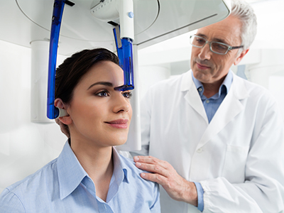
A CBCT appointment is typically quick and uncomplicated. Before the scan, the dental team will review your medical and dental history and remove any removable metal objects—such as eyeglasses, jewelry, or removable dentures—that could interfere with image quality. The procedure is noninvasive and performed with the patient either sitting or standing, depending on the unit design.
During the scan, you will be asked to remain still for a brief period while the machine rotates around your head. The exposure itself usually lasts only a few seconds to a minute, and the total visit time is often less than 15 minutes. There is no discomfort associated with the scan, and most patients find the process straightforward and quick.
Once images are acquired, our team reviews the data and uses specialized software to analyze the anatomy and plan treatment. In many cases, clinicians can show patients the 3D images and explain findings visually, which helps patients understand the proposed care and the rationale behind specific recommendations.
CBCT has become an essential diagnostic resource in contemporary dental practice because it enhances diagnostic clarity, improves treatment planning, and supports safer, more predictable care. If you have questions about whether CBCT is recommended for your situation or would like to learn how three-dimensional imaging could benefit your treatment plan, please contact our office for more information.


Cone-beam computed tomography, commonly called CBCT, is a three-dimensional dental imaging modality that captures volumetric data by rotating an X-ray source and detector around the patient’s head. The system collects multiple projection images and reconstructs them into a 3D model that clinicians can view in axial, coronal, and sagittal planes. This volumetric view reveals the position and relationship of teeth, bone, nerves, and sinuses with a level of anatomical detail that exceeds conventional two-dimensional radiographs.
CBCT is a diagnostic platform rather than a treatment, and it supports many areas of dental care by improving visualization and measurement. Because the data are volumetric, clinicians can take precise linear and angular measurements, assess bone thickness, and inspect root anatomy before choosing a treatment pathway. The clarity and flexibility of CBCT imagery make it a foundational tool for complex diagnostics and treatment planning.
Traditional dental X-rays create two-dimensional images that flatten three-dimensional structures, which can obscure overlapping anatomy and limit certain measurements. CBCT, by contrast, produces cross-sectional and volumetric images that allow clinicians to inspect structures layer by layer and view anatomy from multiple angles. This 3D perspective reduces ambiguity when evaluating root morphology, bone contours, and the location of critical structures such as the inferior alveolar nerve.
Because CBCT captures a volume rather than a single plane, it can reveal findings that would be missed or uncertain on standard films, such as localized bone defects, hidden root fractures, or complex canal configurations. The improved diagnostic confidence from CBCT often leads to more targeted treatment plans and fewer intraoperative surprises.
Clinicians commonly recommend CBCT for situations that require high-resolution spatial information, including dental implant planning, assessment of impacted or unerupted teeth, evaluation of complex endodontic cases, and preoperative planning for extractions or bone grafting. CBCT is also useful for orthodontic assessment when skeletal relationships or airway considerations are important to the treatment plan. The decision to use CBCT is made on a case-by-case basis and depends on the diagnostic question at hand.
Dental teams follow the principle of using the smallest field of view and lowest exposure that will answer the clinical question, so not every patient or every visit requires CBCT. Pregnant patients and those with specific radiation concerns should discuss timing and alternatives with the clinician, and the team will consider deferral or other imaging options when appropriate. Ultimately, CBCT is recommended when the expected diagnostic benefit outweighs the minimal radiation exposure.
Successful implant therapy depends on accurate assessment of bone volume, bone density, and the spatial relationship between proposed implant sites and nearby anatomical structures such as nerves and sinuses. CBCT provides precise three-dimensional measurements that enable clinicians to select the ideal implant length, diameter, and angulation before surgery. With volumetric data, clinicians can simulate implant placement virtually and evaluate whether bone grafting or sinus augmentation will be necessary.
When indicated, CBCT data can be used to fabricate surgical guides that translate the digital plan to the operative field, which reduces intraoperative guesswork and shortens chair time. This workflow increases surgical predictability and contributes to better long-term outcomes by optimizing implant position and primary stability. The combined use of CBCT and digital planning supports a more controlled and efficient implant process.
Modern dental CBCT units are designed to limit radiation exposure by using focused fields of view and adjustable exposure parameters tailored to the diagnostic need and patient size. In general, dental CBCT delivers a substantially lower radiation dose than most medical CT scans while providing the higher-resolution images required for dental diagnostics. Clinicians follow the ALARA principle—keeping exposure as low as reasonably achievable—when determining whether to recommend a scan.
Protective measures and equipment positioning are used as appropriate to minimize exposure, and alternative imaging or deferral may be considered for patients with heightened sensitivity to radiation. Pregnant patients should always inform the dental team so that individualized precautions or scheduling decisions can be made. The final decision balances diagnostic benefit, patient safety, and clinical necessity.
A typical CBCT appointment is brief and noninvasive, often lasting less than 15 minutes from check-in to completion of imaging. Before the scan, the clinical team will review your medical and dental history and ask you to remove removable metal objects such as glasses, jewelry, or dentures that could create image artifacts. Depending on the unit, you may be seated or standing and will be asked to remain still while the scanner rotates around your head.
The actual X-ray exposure commonly lasts only a few seconds to under a minute, and you should experience no discomfort during the procedure. After images are acquired, the clinician uses specialized software to reconstruct and analyze the volume and will often review the findings with you, explaining how the images inform diagnosis and treatment planning. This visual review helps patients understand proposed care and the rationale behind recommended steps.
Yes, CBCT can provide valuable information about nearby structures such as the paranasal sinuses, nasal cavity, airway passages, and temporomandibular joints when those areas fall within the scanned field of view. The cross-sectional images help clinicians identify sinus pathology, airway constriction, or bony changes in the jaw joint that may influence dental or medical treatment. Because CBCT offers a 3D perspective, it can reveal the true extent of anatomic relationships that are important to both dental and interdisciplinary care.
While CBCT is a useful tool for screening and planning, it is not always a substitute for medical imaging when soft tissue resolution or larger regional assessment is required. When findings suggest a clinically significant airway or sinus disorder, the dental team may recommend collaboration with a medical specialist for further evaluation and comprehensive management. CBCT serves as an important bridge between dental diagnosis and broader medical assessment when indicated.
In endodontics, CBCT helps identify complex root canal anatomy, accessory canals, root fractures, and periapical lesions that may be difficult to detect on standard radiographs. The additional detail guides decisions about nonsurgical retreatment, the need for surgical endodontic procedures, or targeted interventions that preserve tooth structure. By clarifying the extent and nature of pathology, CBCT supports more accurate diagnoses and more effective treatment planning.
For restorative and prosthetic care, CBCT informs assessments of bone support, crown and bridge planning, and full-mouth rehabilitation by revealing true root positions and bone contours. The images help clinicians design restorations that respect underlying anatomy and prosthetic requirements, improving fit and long-term function. Integrating CBCT with intraoral scans and clinical examination yields a more comprehensive foundation for restorative decisions.
While CBCT excels at showing hard tissue details, it provides limited soft tissue contrast compared with medical CT or MRI, so it is not optimal for evaluating soft tissue pathology alone. Metal restorations, implants, or orthodontic appliances can produce beam-hardening artifacts that obscure adjacent structures and reduce diagnostic clarity in localized areas. Additionally, CBCT is not indicated for routine screening in every situation and should be reserved for cases where the added 3D information will directly influence clinical decision-making.
Another consideration is that accurate interpretation requires training and experience, so clinicians must integrate CBCT findings with the patient’s history, clinical exam, and other diagnostic records. Field-of-view selection is important; a larger field captures more anatomy but may increase exposure, so clinicians balance diagnostic needs against exposure principles. Recognizing these limitations ensures CBCT is used appropriately and effectively.
At the office of Schneider Family & Cosmetic Dentistry in Mooresville, CBCT is incorporated into care when three-dimensional information will materially improve diagnosis, surgical planning, or restorative design. The team uses volumetric data to plan implant placement, evaluate complex endodontic cases, assess impacted teeth, and support orthodontic or airway evaluations, matching the field of view and exposure settings to each clinical question. This targeted approach helps minimize surprises during treatment and supports more predictable clinical results.
Clinicians at the practice review CBCT images with patients using visualization software so individuals can see the anatomy and understand the rationale for recommended care. By combining CBCT data with clinical examination and digital workflows, the team tailors treatment plans, improves surgical safety, and coordinates interdisciplinary care when needed. Patient safety and diagnostic value guide every decision about imaging, ensuring CBCT is used responsibly to enhance outcomes.

Ready to schedule your next dental appointment or have questions about our services?
Contacting Schneider Family & Cosmetic Dentistry is easy! Our friendly staff is available to assist you with scheduling appointments, answering inquiries about treatment options, and addressing any concerns you may have. Whether you prefer to give us a call, send us an email, or fill out our convenient online contact form, we're here to help. Don't wait to take the first step towards achieving the smile of your dreams – reach out to us today and discover the difference personalized dental care can make.